CONCEPTS OF STRUCTURE CLASSIFICATION OF RESPIRATORY INTERNAL ORGANS
In fact, there are many parts of the respiration organ such as an upper and lower respiratory tracts including the airways, the lungs and linked blood vessels, and the muscles that enable breathing. The lower respiratory tract or lower airway is derived from the developing foregut and it consists of the trachea, the bronchi and bronchioles, and lungs including alveoli. The upper respiratory tract includes the organs located outside of the chest cavity area such as nose, pharynx, larynx, whereas, the lower respiratory tract includes the organs located almost entirely within it (e.g. trachea, bronchi, bronchiole, alveolar duct, alveoli, and diaphragm). Therefore, this standard will provide the structure of (1) upper respiratory tract, (2) lower respiratory tract, and (3) hierarchy of its structure.

Figure1: A complete, schematic view of the human respiratory system with their parts and functions
Hierarchical structure is an arrangement of items (objects, names, values, categories, and so on) in which the items are represented as being “above”, “below”, or “at the same level as” one another. The hierarchical structure is based on the skeletal figure which consists of a tree of objects that defines the transformations from root to the end effector of each appendage of the humanoid. In computer science, a hierarchical tree structure is widely-used with a set of linked nodes. By defining the section point as the node of a tree, we can get a clear context between the section points or branching structures. Therefore, the hierarchical structure is useful for understandings of the representing 3D respiratory internal organ. It is created based on the anatomy object models included lower and upper respiratory tracts by using nodes and edges.
Figure 2, The sets of nodes and edges are separated by hierarchical category’s names. They are combined together into skeletal internal organ represents for the 3D respiratory internal organ of human beings. The skeletons of human internal organ system are defined to make a connection for creating animation of each respiratory organ. In the respiratory internal organ animation’s processes, air enters a human body through the nostril. It moves through the throat and then the trachea. From the trachea, it enters the bronchi and then goes into the lungs. The bronchi form a network of tubes known as bronchioles. Each Hemoglobin absorbs oxygen from the air in the lungs and carries it to tissues all over the body.
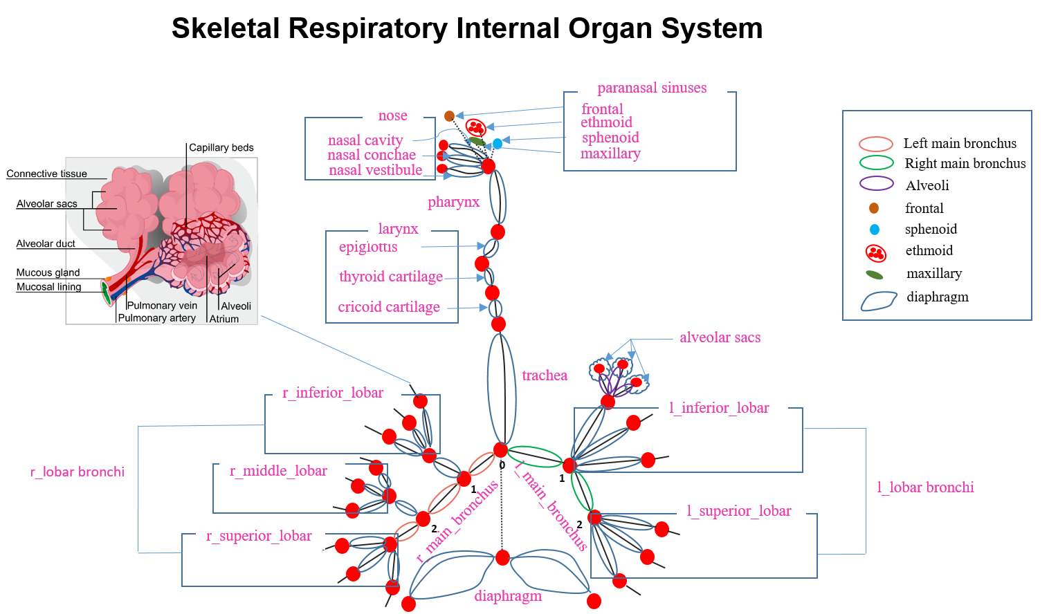
Figure2: The skeletal respiratory internal organ system connection with nodes and edges to represent to 3D respiratory system of human beings
Figure 3 shows a whole respiratory internal organ tree structure for human beings from diaphragm to paranasal sinuses. Diaphragm contains a central tendon, vena caval foramen, esophageal hiatus, diaphragm, right crus, and left crus. The structure of trachea and right main bronchus consist of trachea, right main bronchus, right inferior lobar, right middle lobar, and right superior lobar; whereas, trachea, left main bronchus, left inferior lobar, and left superior lobar are parts located in the left side. There are also right and left lungs category component’s names which are apex of lung, superior lobe, horizontal fissure, middle lobe, oblique fissure, and inferior lobe. Larynx is located between pharynx and trachea. It has epiglottis, thyrohyoid membrane, thyroid cartilage, cricothyroid ligament, cricoid cartilage, and thyroid gland. Furthermore, pharynx, nose and paranasal sinuses inhere in many organ’s parts as shown in the figure image above.
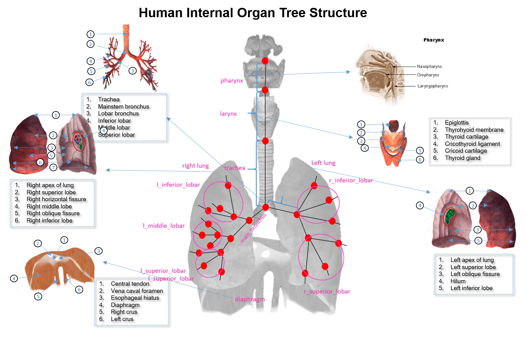
Figure3: Internal organ structure of human beings from diaphragm to paranasal sinuses